
Presentation
Headache, vertigo and seizures
Patient Data
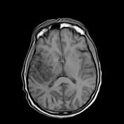

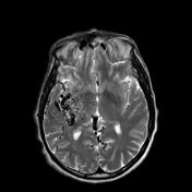
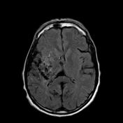
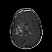
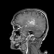
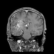

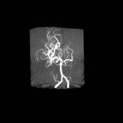
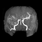

Large nidus of malformed vessels with serpigenous appearance (bag of worm) seen involving the right superior temporal and inferior frontal regions. Although large no mass effect could be detected. Arterial supply derived from the right posterior and middle cerebral arteries. venous drainage to the cortical veins and right internal cerebral vein. No hemorrhage could be detected. It displays flow void signal in T2 and SWI with homogenous contrast enhancement after contrast administration.
Case Discussion
Cerebral AVM may be parenchymal, dural or mixed according ot its arterial supply and venous drainage. The hallmark of the disease is the presence of nidus which is a large collection of arteries and veins that communicate with each others without capillary bed though arteriovenous shunting occur.


 Unable to process the form. Check for errors and try again.