
Presentation
Skin mass in left lower abdomen.
Patient Data
Age: 60 years
Gender: Male
From the case:
Dermatofibrosarcoma protuberans

Download
Info

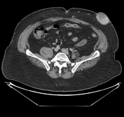
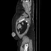
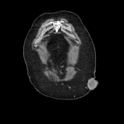
CECT shows well define enhancing dermal / subcutaneous mass with small areas of necrosis within left anterolateral abdominal wall without underlying invasion of abdominal wall muscles. There were no signs of metastasis within chest or abdomen.
Case Discussion
Histopathological diagnosis: dermatofibrosarcoma protuberance.


 Unable to process the form. Check for errors and try again.