Presentation
An adult female complains of right iliac fossa pain.
Patient Data
Age: 40 years
Gender: Female
From the case:
Appendiceal mucocoele




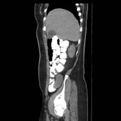

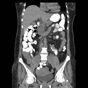


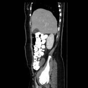

Download
Info
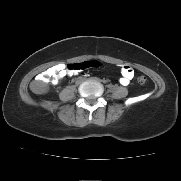
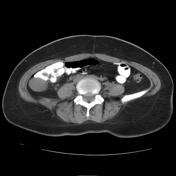
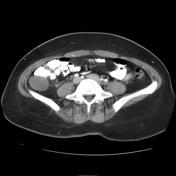
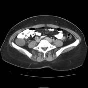
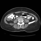
An oblong thin walled, cystic mass near tip of the cecum. It measures about 7.7 x 3 cm. It has homogenous hypodensity ( fluid density ) in pre contrast study. It shows faint peripheral wall enhancement in delayed phase after IV contrast with dirty mesenteric fat at its base. Finding mostly representing appendiceal mucocoele.
Case Discussion
A tubular cystic structure is seen at the right iliac fossa. It lies in intimate relation to the cecum. On top of the differential is an appendiceal mucocele, which was proven postoperatively.


 Unable to process the form. Check for errors and try again.