
Presentation
Back pain and constitutional symptoms.
Patient Data

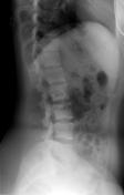


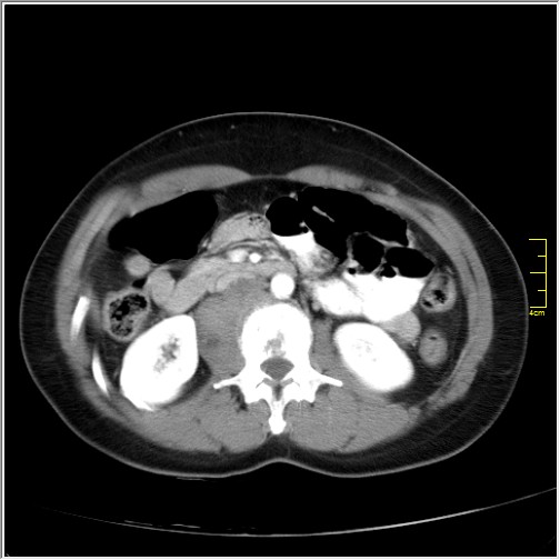
A little over one year later, there is a poorly defined, mixed lytic and blastic lesion within the right lateral aspect of the L2 vertebral body. An associated heterogeneous soft-tissue mass extends from this lesion into the right retroperitoneal region inferior to the right renal artery.


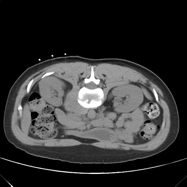
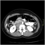
Surface markers were placed over the right paraspinal region to the determine biopsy entry point. The medial marker and proper slice position were selected for optimal tumor access. A slightly medially oriented needle trajectory was used to avoid the kidney and obtain an adequate central core sample from the paraspinal mass.

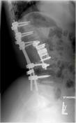
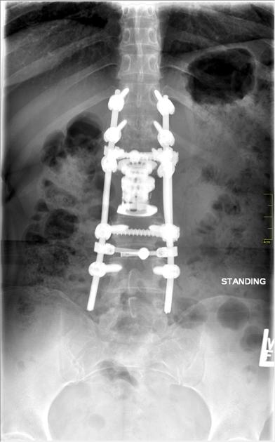
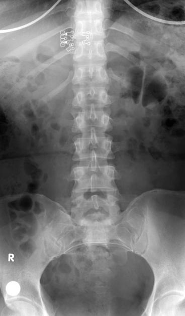
The L2 vertebra and associated tumor has been resected, with replacement by an expandable titanium cage. The region is stabilized with bilateral pedical screw fixation from T11-L4 and anterior transverse fixation at the levels above and below L2.
Case Discussion
Ewing sarcoma is a rare tumor in those older than 25 years of age (95% affected are between 4 and 25 years of age), and also uncommonly involves the axial skeleton (~15% involve axis). In this case, the patient is both unusually old and affected in an uncommon location.
Approximately 40% of Ewing tumors show some amount of sclerosis, but sclerosis is more common in affected flat bones than in long bones, and is a feature in this case.
The differential diagnosis in this case includes: metastatic lesion (breast, gynecologic, etc), lymphoma, myeloma, chordoma, osteosarcoma, and other unlikely primary bone tumors.


 Unable to process the form. Check for errors and try again.