
Presentation
Mass on chest X-Ray.
Patient Data
Age: 72 years
Gender: Male


Download
Info

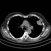
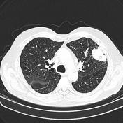
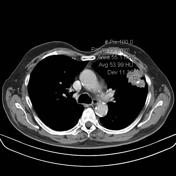
Solid well-demarcated mass with irregular contours in the left upper lobe with retraction of the adjacent pleural surface. There is a small calcified nodule within the mass, probably corresponding to a granulomatous lesion engulfed by the tumor. There is contrast medium enhancement.
Case Discussion
Ultrasound guided percutaneous biopsy diagnosed squamous cell carcinoma of lung origin.
This cell type accounts for ~30-35% of all lung cancers and usually occurs in heavy smokers. Although squamous cell carcinoma of the lung is traditionally a central tumor (66-90%), the incidence of peripheral tumors is increasing and can lead to chest wall invasion.


 Unable to process the form. Check for errors and try again.