
Presentation
Recurrent chest infections, chronic productive cough.
Patient Data
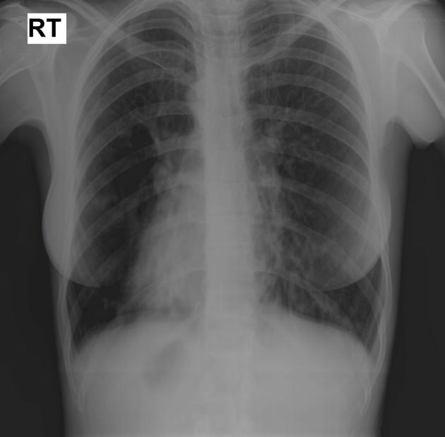
Right sided heart and gastric gas bubble with linear shadows seen in both lungs (more evident at lower zones) suggestive of bronchiectasis.
Relative hyperinflated lungs.
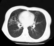


Situs inversus (right-sided heart, aorta, stomach, spleen and left-sided liver).
Bronchiectasis - tree in bud appearance (suggestive of superadded infection).
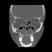

Bilateral maxillary and ethmoidal sinusitis, as well as sphenoidal sinusitis.
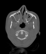
Bilateral otomastoiditis.
Note: Ciliated epithelium covers most areas of the upper respiratory tract, including the nasal mucosa, paranasal sinuses, middle ear, eustachian tube, and pharynx. The lower respiratory tract contains ciliated epithelium from the trachea to the respiratory bronchioles.
Case Discussion
Typical case of Kartagener syndrome, including chest x-ray and CT studies as well as CT scanning of the paranasal sinuses.
The triad of situs inversus, bronchiectasis and sinusitis is clearly shown in this case.


 Unable to process the form. Check for errors and try again.