
Presentation
Incidental finding
Patient Data


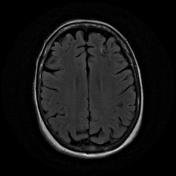
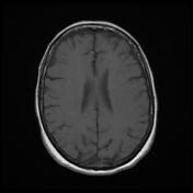
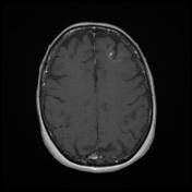
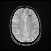
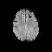
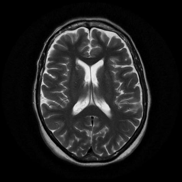
Within the anterior aspect of the left frontal lobe, are typical features of a developmental venous anomaly with associated hemosiderin staining suggestive of a cavernoma. The only enhancement visible views of the DVA. A 2nd smaller region of susceptibility induce signal loss is noted in the very anterior aspect of the superior frontal gyrus, presumably representing a 2nd smaller cavernous malformation.
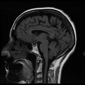
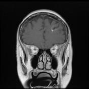
The remainder of the brain is unremarkable in appearance. MRA demonstrates no abnormal vessels in the region (unfortunately the top of the MRA only barely includes the lesion), and no abnormal flow voids are seen on T2-weighted sequences. The remainder intracranial circulation is unremarkable with no stenoses, abnormal vessels, or aneurysm is identified.
Case Discussion
Typical appearances of DVA and cavernoma, frequently co-existing.


 Unable to process the form. Check for errors and try again.