
Presentation
Three days of bowels not opening and increasing abdominal distension.
Patient Data

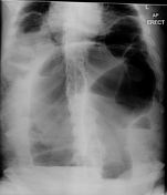
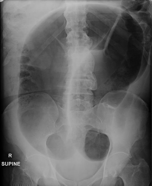
Grossly-dilated loop of large bowel has a 'coffee-bean shape' and the descending colon tapers in its inferior portion in keeping with a sigmoid volvulus. Normally positioned cecum and ascending colon. Air-fluid levels on erect projection.


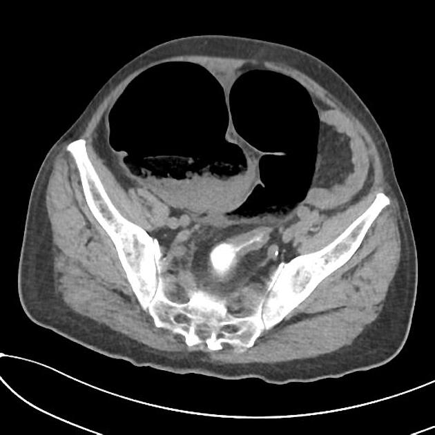
Grossly dilated loop of sigmoid colon with whirl sign of the sigmoid mesentery. Fat stranding of the sigmoid mesentery. No mural thickening appreciated. Remainder of the colon and small bowel is unremarkable.
Liver, spleen and pancreas have a normal appearance as for the patient's age and non-contrast study. Cholecystectomy clips. Atrophic kidneys with 1.7 cm hypodensity and 1.1 cm exophytic hypodensity at the left interpolar region. Small volume of perisplenic free fluid. No free gas. Lung bases are clear. Degenerative lumbar spine changes.
Case Discussion
The large central lower-abdominal gas-filled viscus represents the distended sigmoid colon that has volved at its mesenteric base causing obstruction. On the coronal reformat of the CT, the swirling pattern of the vessels at the base of the mesentery is seen and confirms the cause of sigmoid distension.
Features here are of sigmoid volvulus.


 Unable to process the form. Check for errors and try again.