
Presentation
History of V/P shunt placement many years ago. Now presents with abdominal distension.
Patient Data
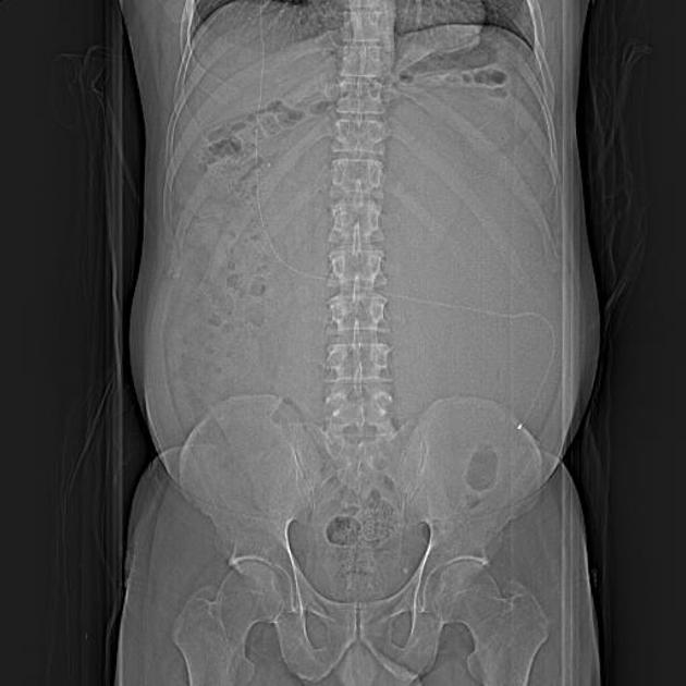
The ventriculoperitoneal shunt is travesring the abdomen at the scout image


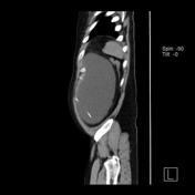
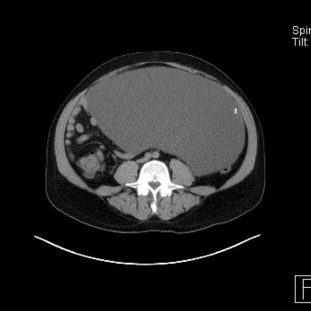
Huge intra-abdominal cyst is surrounding the termination of the venticuloperitoneal shunt measuring 26.9 x 17.4 x 36.1 cm along its maximum transverse, anteroposterior and craniocaudal dimensions.
Case Discussion
A peritoneal CSF pseudocyst is a rare complication of ventriculoperitoneal shunt catheter placement. The time from the last shunting procedure to the development of an abdominal pseudocyst ranges from 3 weeks to 5 years.
CT
May show a small or massive , loculated cyst like structure in the peritoneal cavity at the distal tip of VP shunt
Measurement of attenuation values with CT characterizes the contents as water attenuation and demonstrates the relationships of portions of the shunt catheter with the pseudocyst.


 Unable to process the form. Check for errors and try again.