
Presentation
Middle aged female with a short history of headache and lethargy. No medical history.
Patient Data
Age: 50
Gender: Female
From the case:
Cerebral arteriovenous malformation - ruptured

Download
Info

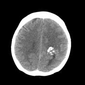
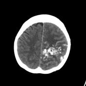
Left parietal lobe focal calcification.
Acute intraventricular hemorrhage.
Left MCA and PCA feeder arteries.
Draining vein into superior sagittal sinus.
Early hydrocephalus.
Incidental basilar tip aneuerysm.
From the case:
Cerebral arteriovenous malformation - ruptured

Download
Info
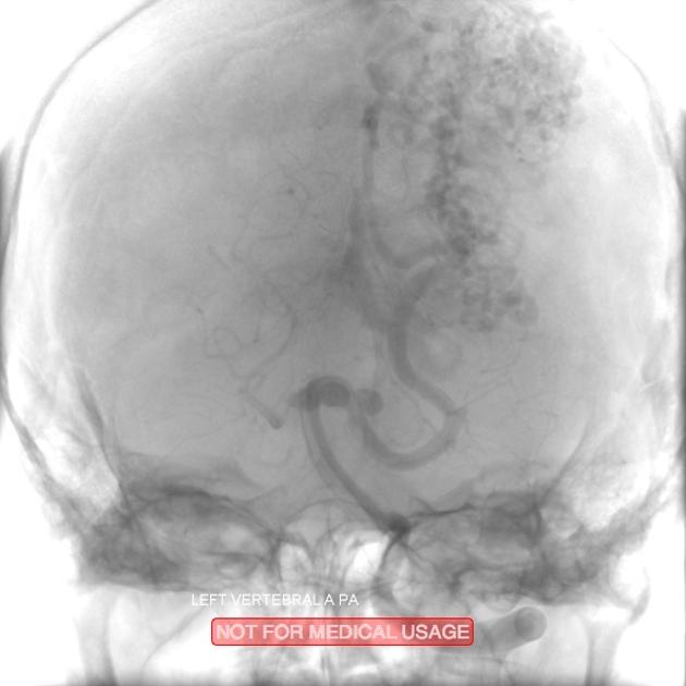
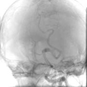
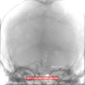
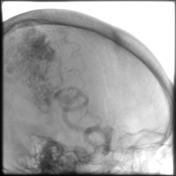
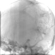
Posterior and middle cerebral feeder vessels into a large AVM.
Huge left parietal AVM is shown as a 'mesh' of serpigenous vessels
Case Discussion
This large cerebral AVM presented following intra-ventricular hemorrhage.
The gold standard catheter angiography, correlates well with the CT findings of feeder vessels from the posterior and middle cerebral arteries, with draining into the superior sagittal sinus.


 Unable to process the form. Check for errors and try again.