
Presentation
Recurring carcinoid tumor.
Patient Data

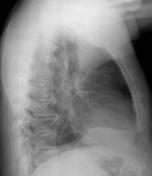
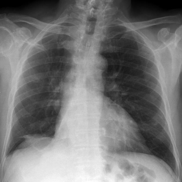
Chest X-ray demonstrates collapse of the righter lower lobe with increase opacity in the medial base of the right lung with lose of medial aspect of silhouette of right hemidiaphragm on both PA and lateral projections. There is shift of mediastinum and trachea to the right and elevation of the right hemidiaphragm


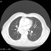
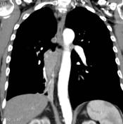
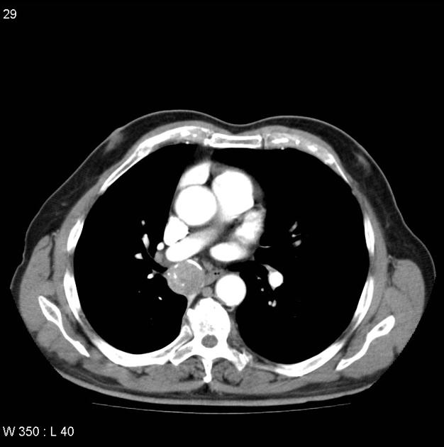
There is a mass lesion located below the right main bronchus which is causing partial occlusion of the right main bronchus. The lesion is heterogeneous in nature with hyper-dense spots consistent with calcification. The lesion shows some degree of enhancement following administration of contrast.
Another homogeneous mass lesion with no contrast enhancement locate near the right hilum.
The heterogeneous hyper-density at right lower base with trachea deviate to right indicate collapse of right lower lobe.
Pathohistology
MACROSCOPIC DESCRIPTION
1. Specimen container labeled 'left tracheal biopsy'. A tan tissue fragment, 3 mm.
2. Specimen container labeled '(R) main bronchus tumor'. Three tan tissue fragments, admixed with blood clot, ranging in size from 5-10 mm.
MICROSCOPIC DESCRIPTION
1. Sections show a single fragment of bronchial tissue. This comprises respiratory type epithelium with attached fibrous stroma infiltrated by nests of epithelioid cells. These cells have rounded nuclei, finely divided chromatin, and moderately abundant amphophilic cytoplasm. The appearances are in keeping a carcinoid tumor. There is no mitotic
activity or necrosis to suggest that this is an atypical carcinoid, based on a small sample.
2. Sections show fragments of bronchial tissue, widely infiltrated by tumor nests with similar features to those described for specimen 1. The growth pattern is also partly trabecular. No necrosis is seen. Up to two mitoses per 10 high power fields are identified. Definite evidence to
place the tumor in the atypical carcinoid category is not identified based on these small samples.
DIAGNOSIS
1&2 Left tracheal biopsy and right main bronchus tumor biopsy - recurrent carcinoid tumor.


 Unable to process the form. Check for errors and try again.