
Presentation
Cough and breathlessness
Patient Data
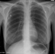

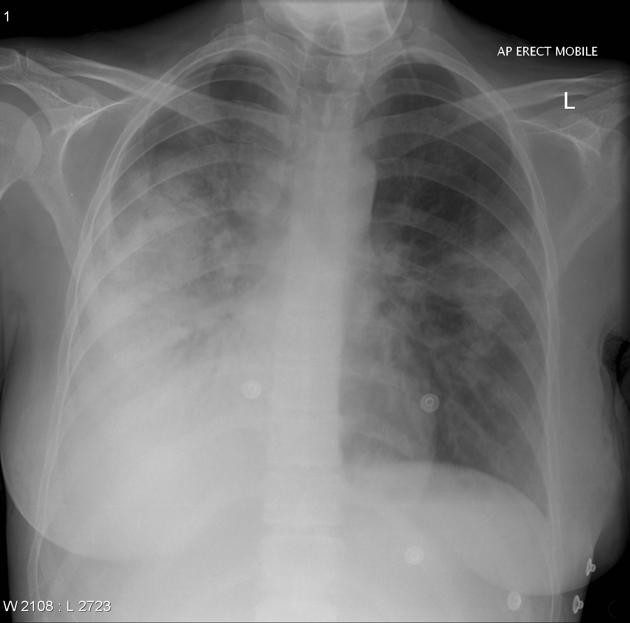
At presentation to ED (vs. 2nd CXR from 5 months' ago) there was widespread opacity across right lung especially lower zone with air bronchograms, indicating consolidation of right lower and/or middle lobes. Some opacity just above horizontal fissure suggesting progression of infection into right upper lobe. Right heart border was lost.
Smaller focal consolidation of the left mid zone.
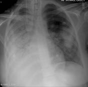
During her admission at hospital, the consolidation spread to right upper lobe and started to develop in left lung with more prominent air bronchogram consistent with widespread infection.

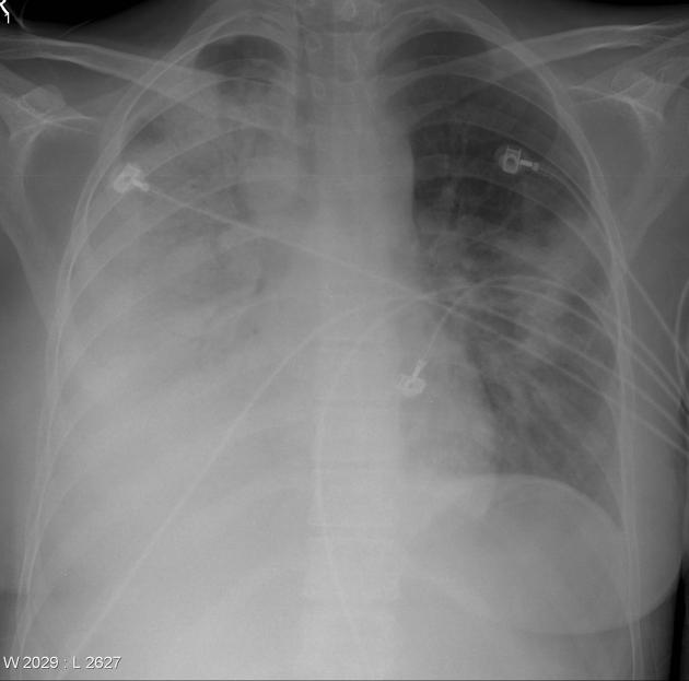
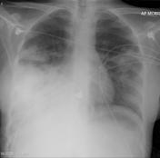
Endotracheal tube and NGT inserted and repeat chest radiograph taken.
Severe right sided consolidation and large focal consolidation of the left mid zone with extension towards lung base.
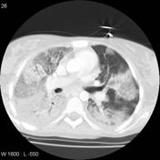

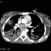
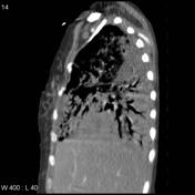
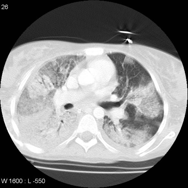
Endotracheal tube and nasogastric tube in situ. Widespread airspace consolidation mixed with ground-glass opacity across both lungs, more severe on right. Very shallow right-sided pleural effusion.
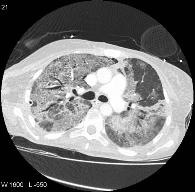
Endotracheal tube in situ.
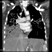
Compared to previous CT, some resolution of consolidation bilaterally but presence of residual ground-glass opacity with fibrotic changes and emphysema which are more obvious in right lung.
Left-sided pleural effusion.
Case Discussion
Streptococcus pneumoniae (= pneumococcus) was isolated in the sputum and blood culture. In bacteremic pneumococcal pneumonia in adults, sputum Gram stain and cultures have sensitivities of 80% and 93%, respectively, provided an adequate specimen is produced prior to therapy. However, in actual clinical practice, sensitivity is lower (< 50%) due to several factors, including an inadequate sputum sample, delayed processing, inability to produce sputum, and prior antimicrobial therapy 2.


 Unable to process the form. Check for errors and try again.