
Presentation
Right pelvic pain and irregular menstruation
Patient Data
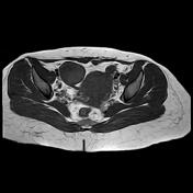


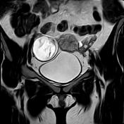
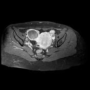
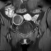


Right ovarian cystic lesion with fluid component hyperintense on T2/STIR and hypointense on T1 and a solid component that is hypointense on T2 and isointense on T1. Enhancement of the cyst wall without the solid component is noted. Large gartner cysts are seen at the anterior vaginal wall.
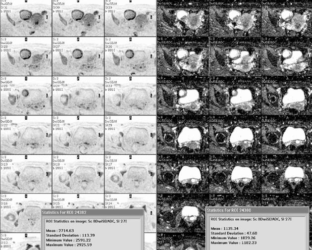
Measuring the ADC values of the solid component and cyst fluid reveals 1.1 and 2.7 x 10⁻³ mm2/s respectively which rules out diffusion restriction.
Case Discussion
Pathological specimen reveals ovarian serous cystadenoma.
DWI can provide an adjunctive tool to differentiate the benign and malignant ovarian lesions. Malignant lesions tend to induce diffusion restriction with ADC value < 0.9 x 10⁻³ mm2/s, the vice versa for the benign lesions. Absence of enhancement of the solid component and absence of diffusion restriction are supportive of benignity.


 Unable to process the form. Check for errors and try again.