
Presentation
Not provided.
Patient Data

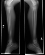
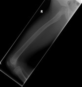
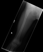

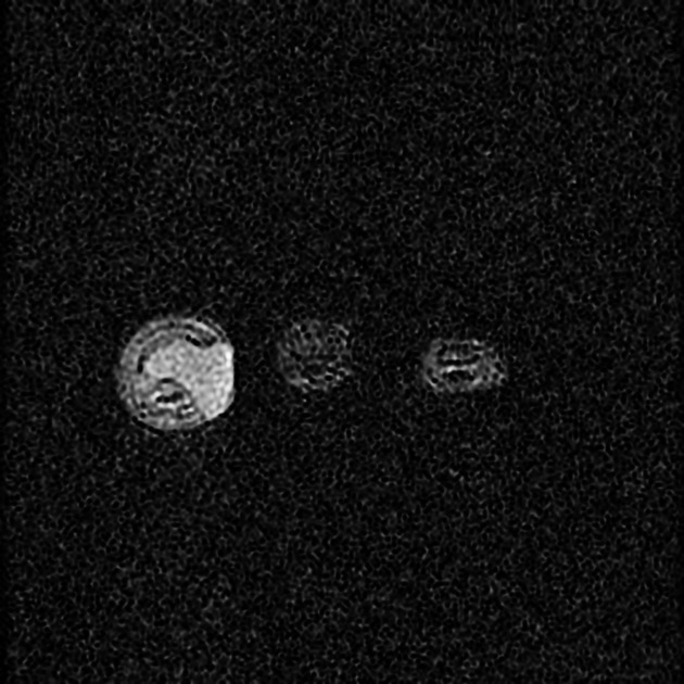
Well-defined lytic lesions are observed in the right medial femoral condyle and calcaneal tuberosities bilaterally.

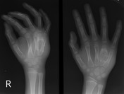
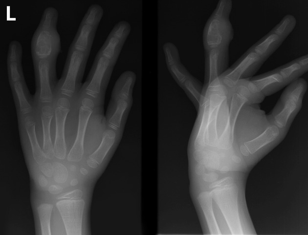
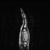
In the Left hand there is an expanslie lytic lesion within the middle 4th phalange that demonstrates cortical violoation along its radial aspect. There is severe surrounding soft tissue swelling, most prominent on the volar aspect, with overlying skin irregularity suggesting an open tract. No periosteal reaction is appreciated.
In the right hand there is an expanslie lytic lesion within the 4th metacarpal that demonstrates cortical violoation along its dorsal aspect. There is severe surrounding soft tissue swelling, most prominent on the dorsal aspect of the hand. No periosteal reaction is appreciated.
Both of these lesions could be described to have a "spina ventosa" appearance.
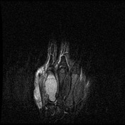

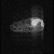
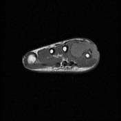

The lesion within the 4th metacarpal of the right hand demonstrates homogeneously high T2 & STIR signal which is isointense to muscle on T1. This lesion extends into the soft tissue through a defect in the dorsomedial cortex of the 4th metacarpal, involving the soft tissue dorsal and medial to the 5th metacarpal, with skin ulceration overlying the abscess on the dorsum and medial aspect of the hand.
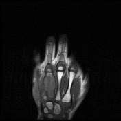
The lesion within the 4th middle phalange of the left hand is similar in appearance to that in the right hand, with homogeneously high STIR signal, extension into the soft tissues,and overlying skin ulceration.
Case Discussion
In patients with coccidioidomycosis, 10-20% will develop granulomatous lesions in the skeleton. Often times, skeletal infection is bilateral and somewhat symmetric, as is the case in this patient. Some authors have described a predilection for bony protuberances with the granulomatous lesions, which is also noted in this case. Lesions are typically well defined, usually without periostitis, sclerosis or seqeuestra formation, which remains true for all of the lesions seen here. 1


 Unable to process the form. Check for errors and try again.