
Presentation
Palpable abdominal mass
Patient Data
Age: 40 years
Gender: Female


Download
Info
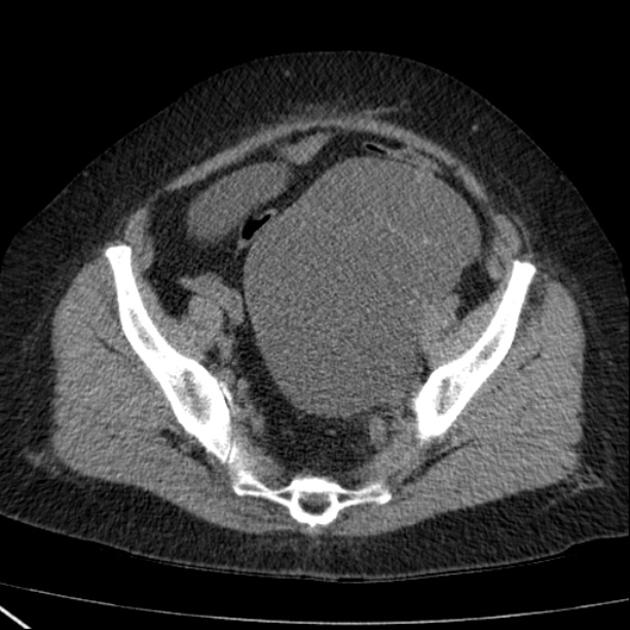
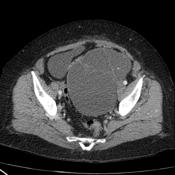
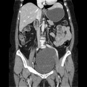
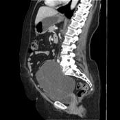
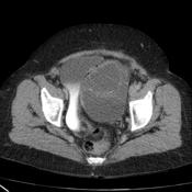
CT of the patient shows a multi-loculated cystic mass adjacent to the bladder, with a smooth
contour, and septa. Small calcifications. The lession shows homogeneous CT attenuation and no endocystic or exocystic vegetation.
Case Discussion
On the basis of this CT the distinction between a serous and mucinous ovarian tumor cannot be made but some features can aid in differentiating mucinous from serous tumors: like a unilocular or multilocular cystic mass with homogeneous CT attenuation , with a thin regular wall or septum, and usually no endocystic or exocystic vegetation is considered to be a serous cystadenoma.


 Unable to process the form. Check for errors and try again.