
Presentation
Chronic cough.
Patient Data
Age: 20
Gender: Female
From the case:
Immotile cilia syndrome

Download
Info
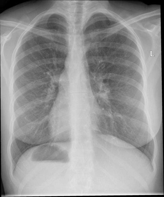
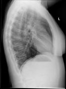
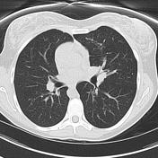
There is dextrocardia. The lungs demonstrate coarsening of the bronchovascular markings, best seen in the lower zones, consistent with bronchiectasis.
From the case:
Immotile cilia syndrome


Download
Info
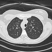
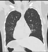
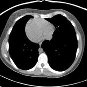
Situs inversus is noted with the aortic arch on the right and the middle lobe on the left. Spleen is also on the right side of the liver on the left.
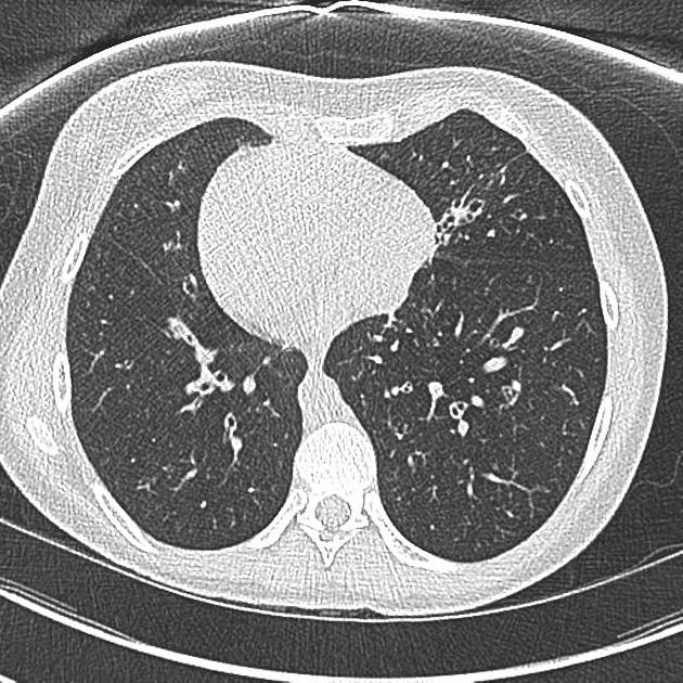
Changes of bronchiectasis are seen in the left middle lobe. There are subtle changes of bronchiectasis in the lingula segment (on the right). No definite bronchiectasis elsewhere. Lungs and pleural cavities are otherwise clear.
No lymph node enlargement is identified.
No suspicious bone lesions identified.
Case Discussion
Typical appearances of immotile cilia syndrome.


 Unable to process the form. Check for errors and try again.