
Presentation
Acute upper back pain.
Patient Data
Age: 50 years
Gender: Male
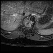
From the case:
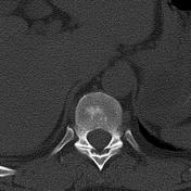
Schmorl node - acute


Download
Info
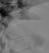
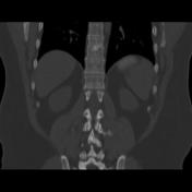
Very subtle intervertebral disc calcifications are seen.
From the case:
Schmorl node - acute
Download
Info

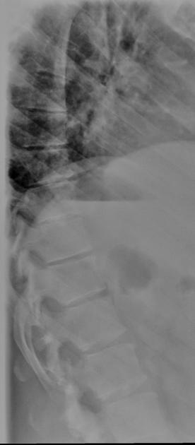
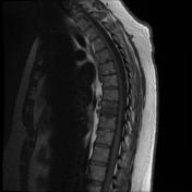
Intervertebral disc calcifications are seen with interruption of the continuity of the superior endplates.
From the case:
Schmorl node - acute

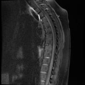

Download
Info

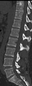
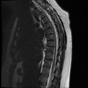
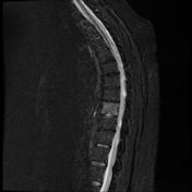
T1 showed reduced bone marrow signal intensity of the vertebral body which appears bright on STIR images in keeping with bone marrow edema. Disc calcification with superior end-plate herniation (Schmorl node) and to a lesser extent the inferior endplate. Findings in keeping with acute Schmorl node.


 Unable to process the form. Check for errors and try again.