
Presentation
Recurrent pneumonia, breathlessness. Previous chest radiograph showed possible diaphragmatic hernia.
Patient Data
Note: This case has been tagged as "legacy" as it no longer meets image preparation and/or other case publication guidelines.
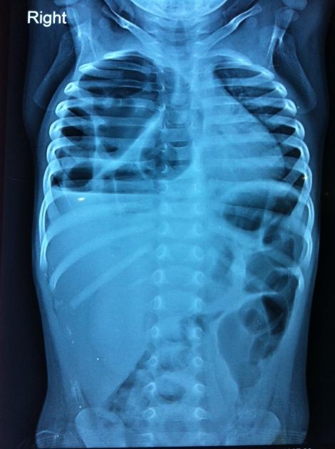
Large cystic areas in right hemithorax with some of them showing air-fluid levels, with contralateral mediastinal shift. Diaphragmatic outline is not well visualized on right side. Possibility of diaphragmatic hernia was being considered.


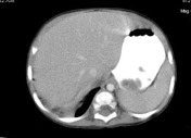
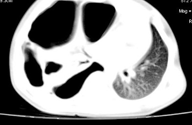
Multiple cystic areas of varying sizes, reaching up to 8 cm in size are seen replacing most of the lung parenchyma. Residual lung parenchyma appears compressed and atelectatic. Contralateral tracheomediastinal shift is seen. Diaphragmatic continuity appears intact.
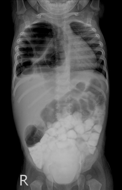
A post-contrast radiograph taken, also shows above mentioned findings.
Case Discussion
Congenital pulmonary airway malformation type I, has large cystic areas, which may have air-fluid levels. Also, these large cysts may produce mass effect and mediastinal shift, which may be confused with congenital diaphragmatic hernia on a radiograph. Cross-sectional imaging helps in this regard.
(Case courtesy: Dr. Abhijit Yadav, Dr. B L Yadav center for diagnostic imaging and research).


 Unable to process the form. Check for errors and try again.