
Presentation
Right proptosis.
Patient Data


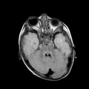
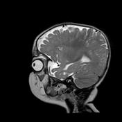
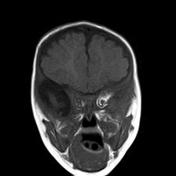
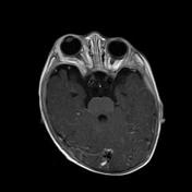
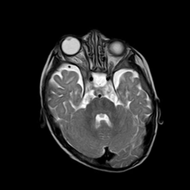
Right sphenoid wing hypoplasia causing deformity of the right orbit which is flattened posteriorly.
Slightly asymmetry dilated CSF spaces at the right middle cranial fossa.
Right upper eyelid subcutaneous soft tissue lesion, that elicits intermediate signal at T1 & T2 WI showing post contrast enhancement.
Right parasellar/cavernous intensely enhancing soft tissue mass lesion is also noted.
The brain parenchyma demonstrates no abnormality.
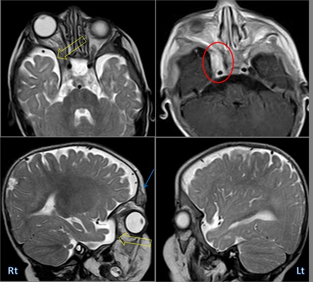
Right sphenoid wing hypoplasia causing deformity of the right orbit which is flattened posteriorly (yellow arrows).
Right parasellar/cavernous intensely enhancing soft tissue mass lesion, possibly, plexiform neurofibroma/schawnnoma (red circle).
Right upper eyelid subcutaneous soft tissue lesion, likely plexigorm neurofibroma (blue arrow).
Case Discussion
Sphenoid wing dysplasia, right orbital plexiform neurofibroma, & cavernous sinus mass that could be; plexiforn neurofibroma or schawnnoma or less likley meningioma (No biopsy was obtained)
Features impressive of Neurofibromatosis type 1,


 Unable to process the form. Check for errors and try again.