
Presentation
CT performed at any alternative institution - clinical indication unknown - reported as a cerebral AVM. MRI to assess further.
Patient Data
Age: 30 years
Gender: Male
From the case:
Cerebral arteriovenous malformation


Download
Info

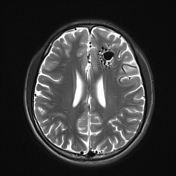
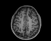
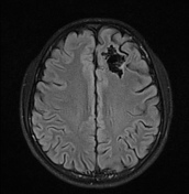
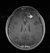
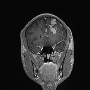
Convoluted vessels in the left frontal lobe with internal flow voids.
Feedings veseels from the anterior and middle cerebral artery derived vessels.
Enlarged cerebral vein draining into the superior sagittal sinus at the convexity.
Case Discussion
Large cerebral arteriovenous malformation.
The key of imaging includes identifying the feeder arteries, draining veins and classifying the AVM.
Catheter angiography is typically performed prior to either radiological or surgical intervention.


 Unable to process the form. Check for errors and try again.