
Presentation
Right upper limb partial seizure.
Patient Data


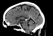
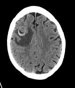
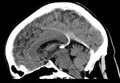
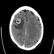
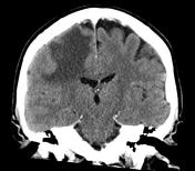
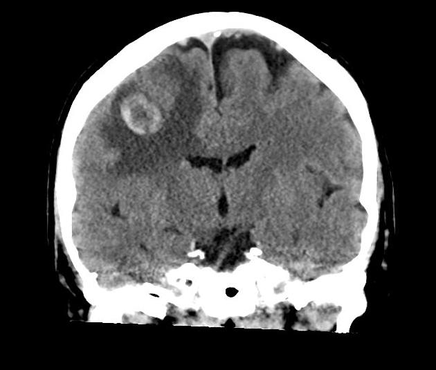
Pre and post contrast scans of the brain have been performed. The 2.2 cm hemorrhagic right frontal mass. There is a moderate amount of adjacent edema with local mass-effect. No other focal abnormality identified.

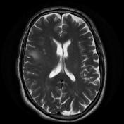
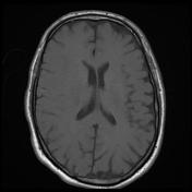
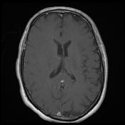
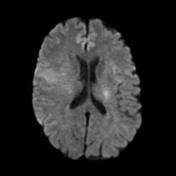
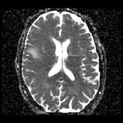

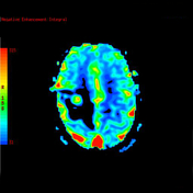
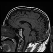
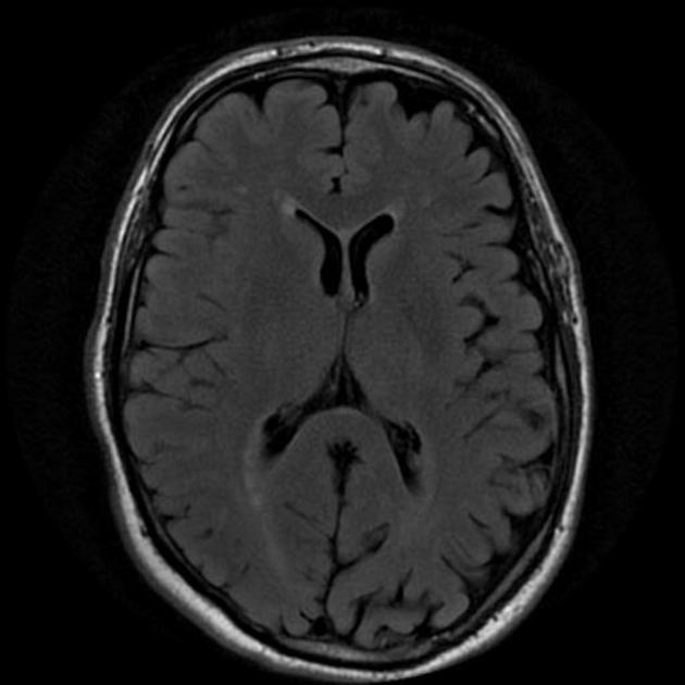
Within the right frontal lobe (precentral gyrus) is a rounded lesion measuring 1.5 cm in diameter with heterogeneous signal including blood products, and vivid heterogeneous contrast enhancement. It is surrounded by a significant amount of vasogenic edema, sparing the cortex. It is an isolated abnormality. Ventricles are unremarkable.
Conclusion:
Right frontal lobe lesion has imaging appearances consistent with a melanoma metastasis or other hemorrhagic metastasis.
Case Discussion
The patient went on to have a craniotomy.
Histology
MICROSCOPIC DESCRIPTION: The section shows fragments of a densely hypercellular tumor and fragments of blood clot and granulation tissue containing collections of hemosiderin filled macrophages. Tumor cells have pleomorphic vesicular nuclei many with conspicuous nucleoli and a variable amount of pale cytoplasm. Finely granular black-brown pigment is noted within the cytoplasm of many tumor cells. There are frequent mitotic figures and foci of tumor necrosis are also noted. Tumor cells show strong immunostaining for tyrosinase. The features are of metastatic malignant melanoma.
FINAL DIAGNOSIS: Metastatic malignant melanoma.


 Unable to process the form. Check for errors and try again.