
Presentation
Enlarged head and hydrocephalus suspected clinically.
Patient Data
Age: 2 months
Gender: Male
From the case:
Alobar holoprosenencephaly


Download
Info
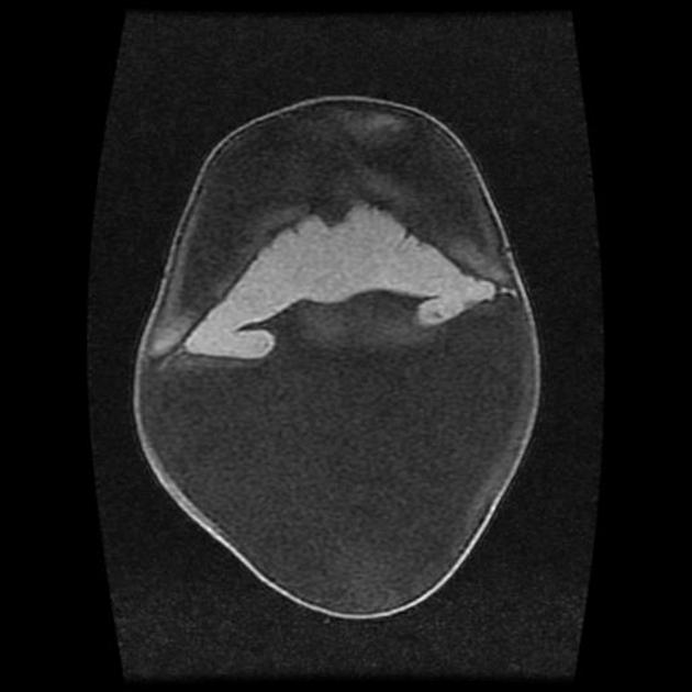
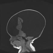
There is absence of normal midline structures and most of the parietal and posterior cortex. Small frontal cortex is present, with no evidence of midline fissure.
The thalami are fused and a large monoventricle and a larger dorsal cyst are present. Cerebellum and brainstem appear within normal limits
Case Discussion
The imaging findings (which were also suspected on the antenatal ultrasound) are typical of alobar holoprosencephaly.


 Unable to process the form. Check for errors and try again.