
Presentation
Severe back pain.
Patient Data


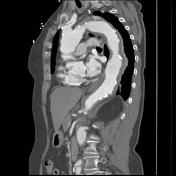
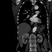
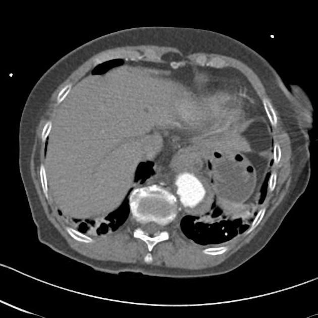
An abnormal appearance of the aortic arch is demonstrated, characterized by a 2 cm amorphous " blob" of contrast present within the dorsal aortic wall approximately 3.5 cm distal to the origin of the left subclavian artery.The descending aorta also has an abnormal appearance, with crescentic wall thickening.
The calcified intima which is atheromatous and irregular is displaced inwardly by this wall thickening. A protuberant atheromatous plaque is noted within the descending aorta.
The intimal displacement and mural thickening extends to the level of the renal arteries.
The arch branches are unaffected.
This appearance, in the current clinical setting is highly suspicious for the presence of an acute intramural hematoma, presumably secondary to active intimal penetration of the aortic arch.
Case Discussion
Key learning points:
- intramural hemorrhage occurs because of rupture of the vasa vasorum
- CT features include hyperdense aortic wall thickening with displacement of the calcified intima often seen


 Unable to process the form. Check for errors and try again.