
Presentation
Swelling of the right front of the head since birth.
Patient Data
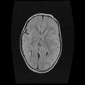

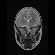
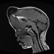

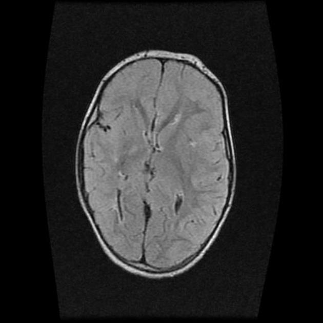
There is a 47mm left frontal cranial defect with herniation of the right frontal lobe, including the anterior horn of the right lateral ventricle. The herniated brain tissue retains high FLAIR signal consistent with gliosis. In addition, there is abnormal signal within the right cingulate gyrus. This is thought to be secondary to compressive ischemia.
There is also associated hypertelorism.
A left frontal developmental venous anomaly (DVA) is also evident.
No hydrocephalus.
Case Discussion
This case demonstrates a large frontal encephalocele, a congenital malformation resulting from a calvarial defect. Other types of encephaloceles include frontoethmoidal encephaloceles, occipital encephaloceles, nasoethmoidal encephaloceles, among others.


 Unable to process the form. Check for errors and try again.