
Presentation
Diffuse pelvic pain and vaginal discharge since two weeks.
Patient Data
Age: 30 years
Gender: Female
From the case:
Tubo-ovarian abscess

Download
Info

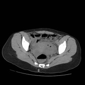
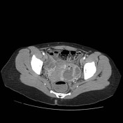
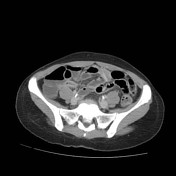
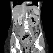
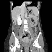
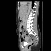
Left sided pelvic parauterine lesion is seen. It has multiple air loculi. It shows marginal enhancement with multilocular appearance in the post contrast study. The surrounding fat planes appear strandy with little peritoneal fluid reaction.The findings are suggestive of pelvic inflammatory disease with abscess formation. An associated small right ovarian cyst is seen.
Case Discussion
Once the patient was diagnosed radiologically she was operated, drained and proved to be tubo-ovarian abscess as a serious complication of pelvic inflammatory disease (PID).


 Unable to process the form. Check for errors and try again.