
Presentation
Abnormal patch on the left iris noticed by the patient.
Patient Data


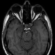
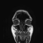
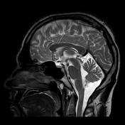
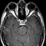

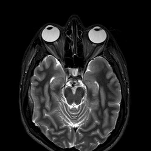
There is a lesion of abnormal signal (increased T1 signal and decreased T2 signal) in the region of the lateral ciliary body & iris of the left orbit, which demonstrates enhancement after gadolinium administration.
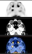

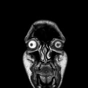
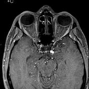
Increased FDG uptake by the fore mentioned left ciliary body lesion (SUV max 4),
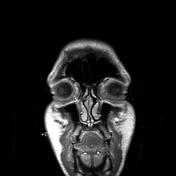
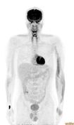
No FDG lesions distinctive for distant metastasis.
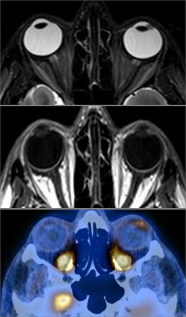
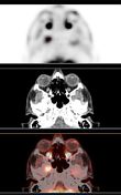
T2, T1+C ,PET/CT
Increased FDG uptake by the fore mentioned left ciliary body & iris lesion (SUV max 4),
Case Discussion
A case of pathologically proven ciliary body and iris melanoma.
Primary uveal malignant melanoma is a cancer (melanoma) involving the iris, ciliary body, or choroid (collectively referred to as the uvea). Tumors arise from the pigment cells (melanocytes) that reside within the uvea giving color to the eye.


 Unable to process the form. Check for errors and try again.