
Presentation
Motor vehicle collision - 60 km/hr.
Patient Data
Age: 40
Gender: Male
From the case:
Supine pneumothorax
Download
Info
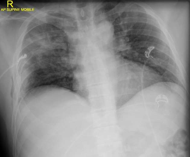
Subcutaneous emphysema is seen in the right lower chest wall along with rib fractures of 4th to 6th right lateral rib fractures.
The right hemidiaphragm has a crisp outline which is suggestive of a pneumothorax.
Right upper zone consolidation.
The left lung is clear. Cardiomediastinal contour is within normal limits for the projection.
From the case:
Supine pneumothorax
Download
Info
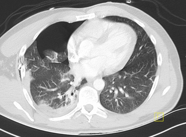
CT confirms rib fractures, subcutaneous emphysema and pneumothorax. Also demonstrates pulmonary consolidation.
Case Discussion
- pneumothoraces on supine chest radiographs ("supine pneumothorax") can be very difficult to pick and often associated factors such as rib fracture and subcutaneous emphysema help raise the suspicion of otherwise equivocal findings
- up to 30% of supine pneumothoraces will be missed
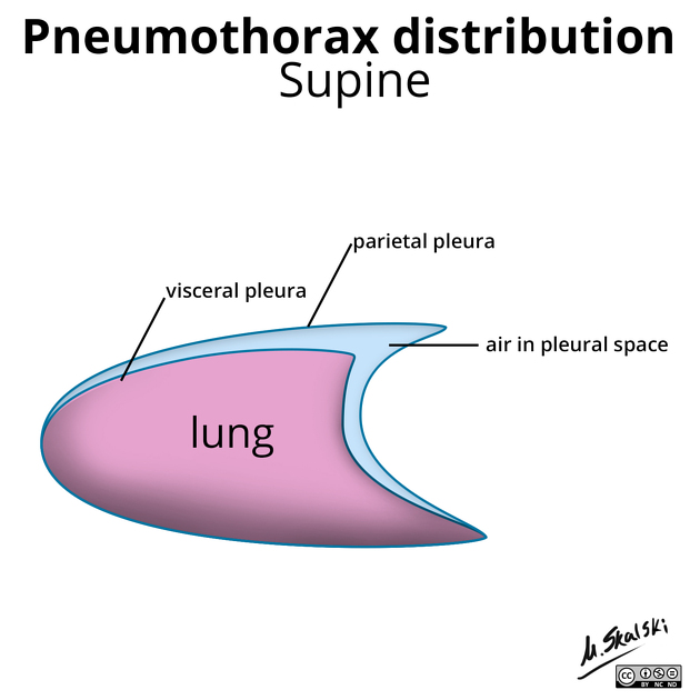
- air collecting in the anteromedial pleural space (as in this case) is the most common location and can result in a dark lucent band that will crisply outline the right heart border and/or right hemidiaphragm
- CT will clearly show a supine pneumothorax


 Unable to process the form. Check for errors and try again.