
Presentation
Left thigh pain and swelling. Plain radiograph reveals aggressive lesion.
Patient Data
Age: 20 years
Gender: Male
From the case:
Ewing sarcoma - femur


Download
Info
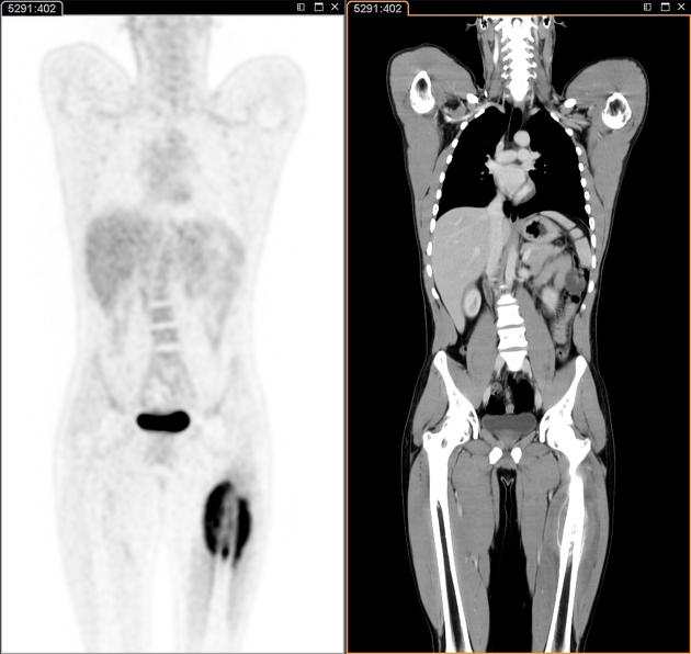
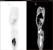
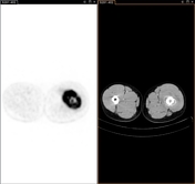
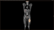
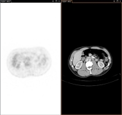
Aggressive predominantly sclerotic marrow lesion of the mid left femoral shaft with associated periosteal elevation and soft tissue extension. PET images reveal avid tracer uptake and delineate surrounding soft tissue extension. No distant metastasis. Mean SUV value ~6.04.
Case Discussion
Biopsy from this lesion revealed Ewing sarcoma. PET-CT was performed to assess tumor extension and search for any distant metastasis.


 Unable to process the form. Check for errors and try again.