
Presentation
Symptoms consistent with a chest infection.
Patient Data
Age: 45
Gender: Female
From the case:
Pleural empyema

Download
Info
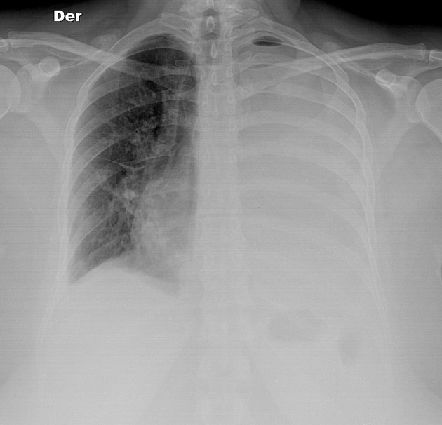
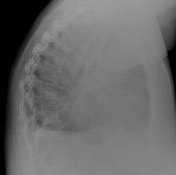
The x-ray shows a large left-side pleural effusion with mediastinal shift. The right lung is clear.
From the case:
Pleural empyema


Download
Info
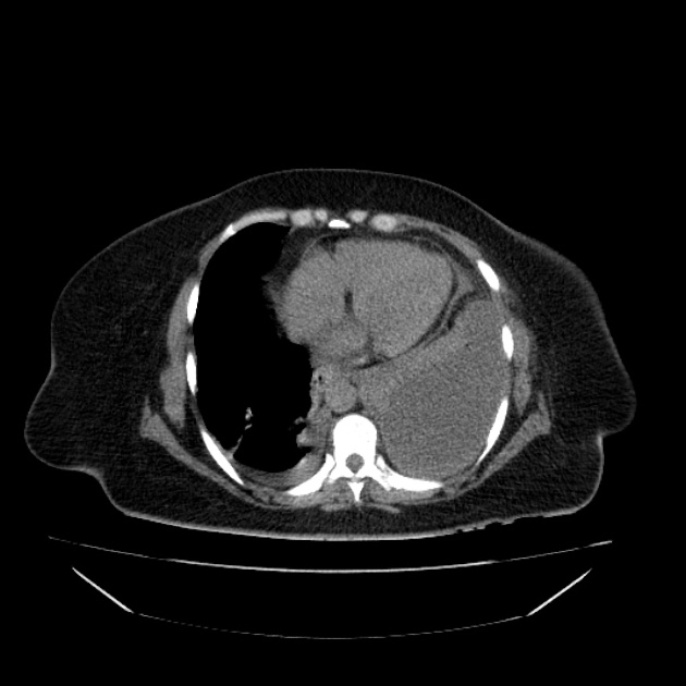
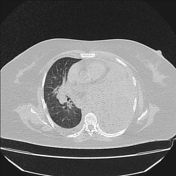
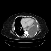
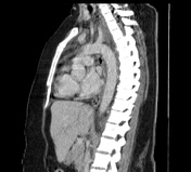
The left hemithorax is filled with a huge pleural collection with small air bubbles inside and with the left lung compressed medially and consequent mediastinal shift.
Case Discussion
This pleural collection was confirmed to be an empyema.


 Unable to process the form. Check for errors and try again.