
Presentation
Febrile convulsion in the setting of upper respiratory tract infection. She was born from noncousin parents, having no remarkable medical history. CT scan underwent for the further evaluation.
Patient Data


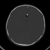
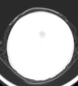
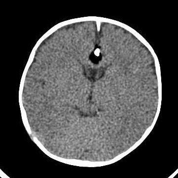
CT scan shows a hypoattenuating sharply demarcated mass lesion associated with calcification which developed in the interhemispheric fissure in the anatomical location of the rostrum and genu of the corpus callosum. There is also partial agenesis of the corpus callosum induced by this mass. There is no evidence of brain swelling.
Case Discussion
Incidental finding of an intracranial lipoma with partial agenesis of the corpus callosum in an infant with febrile seizure. The infant underwent a sepsis workup and was discharged from the hospital in a good situation.
At the first glance, the mass may be misinterpreted as air but a careful radiologist finds it nothing but fat. While intracranial lipoma and pneumocephalus can look identical, but by changing the window or finding the presence of the calcification there, discrimination is possible.


 Unable to process the form. Check for errors and try again.