
Presentation
Headache
Patient Data
Age: 37 yrs
Gender: Male
From the case:
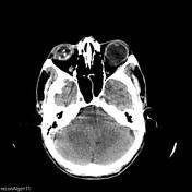
Phthisis bulbi and posterior staphyloma


Download
Info

Right phthisis bulbi and left posterior staphyloma.
Normal study of brain.
Case Discussion
It is important to systematically look at all the structures visible on a CT scan. It is easy to overlook eyeballs when concentrating on brain.


 Unable to process the form. Check for errors and try again.