
Presentation
Known patient with lymphoma after receiving chemotherapy and now for follow up.
Patient Data
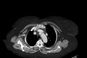


There is a vertical vein that is seen lateral to the aortic arch and ends at the inferior aspect of the left brachiocephalic vein. The left superior pulmonary vein is hypoplastic because this vertical vein drains most of the left upper lobe. Normal appearance of the brachiocephalic veins and SVC. The right atrium is dilated from volume overload.


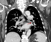
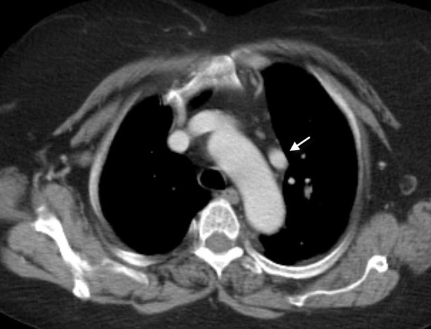
White arrow represents the anomalous vertical vein.
Yellow arrow shows the normal left brachiocephalic vein.
Red arrow refers to the small left superior pulmonary vein.
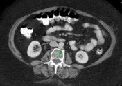
green arrow displays circumaortic left renal vein.
Case Discussion
PAPVR is diversion of portion of the oxygenated blood from the left side of the heart to the right atrium through abnormal connection. This Left to Right shunt elicits volume overload on the right atrium and mixes the oxygenated with non oxygenated blood. Circumaortic left renal vein was another accidental finding. This is not related to the patient's original disease and discovered accidentally.


 Unable to process the form. Check for errors and try again.