
Presentation
Possible seizure, new onset headache.
Patient Data


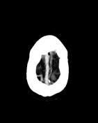
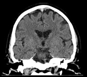
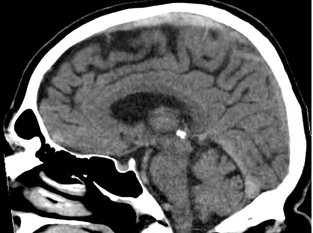
Focal hyperdensity (roughly 70 HU) in the superior sagittal sinus spanning 6-7 cm with surrounding hyperdense cortical veins.
Case Discussion
The patient decided to leave after the study was performed before receiving the results. Despite contacting him by phone and explaining the implications of his diagnosis (venous sinus thrombosis), the patient only returned to the hospital a few days later and went on to suffer several complications despite optimal treatment.
This case demonstrates the importance of systematically checking the dural venous sinuses on CT scans of the brain, especially at its apical portions, seeing as an increased density in this area can easily be missed when looking at axial slices in the brain window. In this case, the hyperdensity is much more clearly seen on the sagittal reconstructions.


 Unable to process the form. Check for errors and try again.