
Presentation
Vertigo
Patient Data
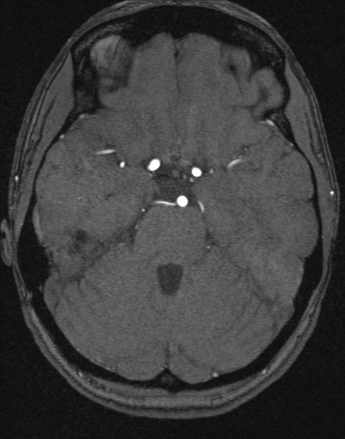
Large right-sided persistent trigeminal artery, from the right ICA to the mid basilar artery is demonstrated.
The basilar artery from this level up to the basilar tip is ectatic. No associated aneurysm.
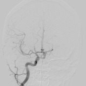

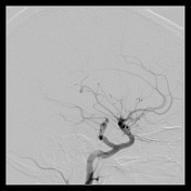
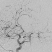
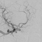
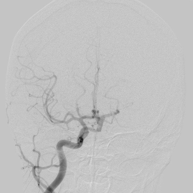
The vertebral arteries are of similar size, with no dissection or focal stenosis from origin to basilar artery. The proximal half of the basilar artery is small caliber, but this is a congenital anomaly secondary to a large persistent trigeminal artery arising from the cavernous portion of the right internal carotid artery. There is fusiform dilatation of the terminal basilar artery, the maximum transverse dimension of 6 mm. There is no saccular, conventional aneurysm. Both carotid arteries are of normal caliber, with no fibromuscular disease or stenosis.
Conclusion
- Large trigeminal artery from right ICA.
- 6 mm fusiform aneurysm of terminal basilar artery-no saccular component, likely low risk, and not likely to benefit from endovascular intervention.
Case Discussion
The persistent primitive trigeminal artery represent the most common form of the carotico-vertebrobasilar anastomoses.


 Unable to process the form. Check for errors and try again.