
Presentation
Surveillance imaging following frequent bleeds.
Patient Data


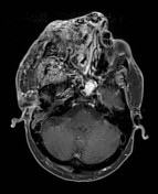
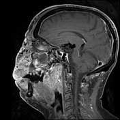

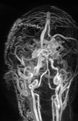
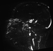

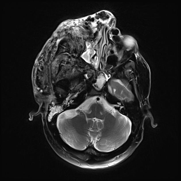
Wyburn-Mason syndrome
Right sided multifocal giant AVM maxillo-facial with involvement of the orbito-nasal space as well as partial involvement of the left side. Involvement of the maxilla, zygomatic bone and the sphenoid. Additional hypothalamic AVM. Soft tissue swelling in the right half of the face, in the parotid, masticator and parapharyngeal space. Dilated vessels on the nose. Exophthalmos on both sides.

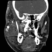
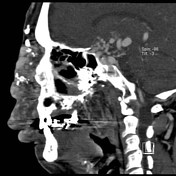
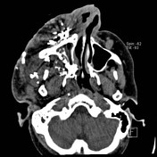
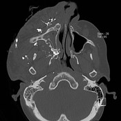
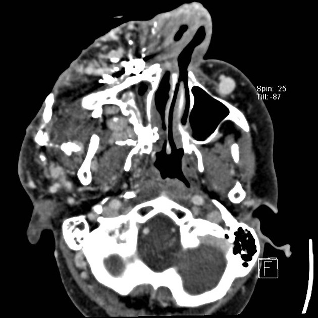
Wyburn-Mason syndrome, corresponding CT (see MRI).
Note hyperdense embolization material on CT images.
Case Discussion
This case illustrates a known case of Wyburn-Mason syndrome, which is part of a wide spectrum of possible phenotypes included in the CAMS - Craniofacial arteriovenous metameric syndrome.
The syndrome was discovered in this patient at the age of 5 years (then not visible externally). Slow growth over years with recurrent bleeding in increasing frequency. Embolization was done several times during lifetime.
The patient ultimately died of large intracranial hemorrhage in his 40s.


 Unable to process the form. Check for errors and try again.