
Presentation
Motor vehicle accident.
Patient Data
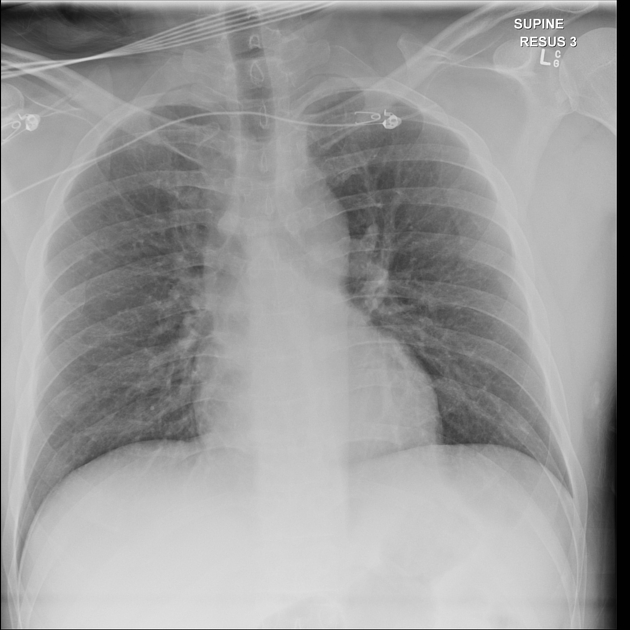
Deep sulcus sign demonstrated on the initial supine chest radiograph which is highly suggestive of a right-sided pneumothorax.
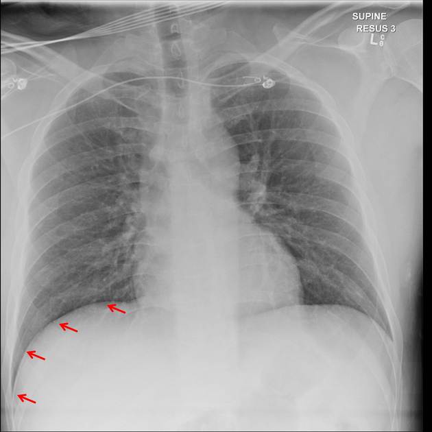
Red arrows labeling the deep sulcus sign on the right.
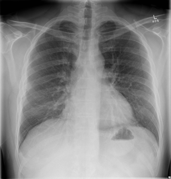
Erect chest radiograph confirming the presence of a small right apical pneumothorax.
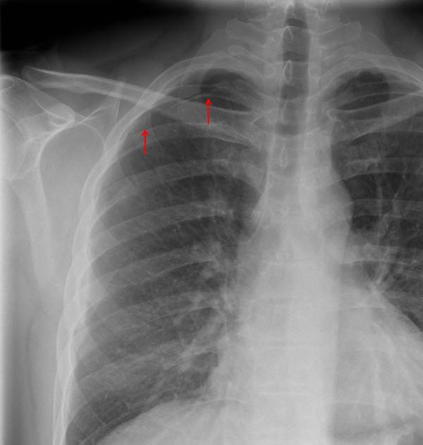
Red arrows labeling the right apical pneumothorax.
Case Discussion
Deep sulcus sign depicts pneumothorax in the supine position as air ascends to the non-dependent pleural space outlining the anterior costophrenic recess/sulcus. If the volume of pneumothorax is sufficiently large it can also outline the costocardic angle and the pericardial fat and additional "pericardial fat pad sign" may also be observed.
A prudent radiologist will need to asses the costophrenic and pericardial region closely when inspecting supine trauma and ICU radiographs as small pneumothorax can be a subtle finding.


 Unable to process the form. Check for errors and try again.