
Note: This case has been tagged as "legacy" as it no longer meets image preparation and/or other case publication guidelines.
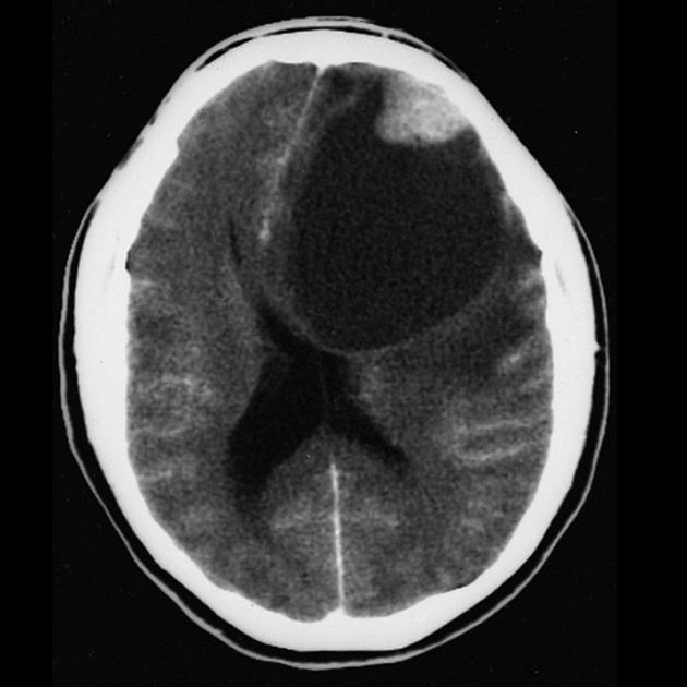
A single image from a contrast enhanced CT of the brain demonstrating a large left frontal lobe cystic lesion with a peripherally located enhancing nodule. This was confirmed to be a pleomorphic xanthoastrocytoma.
Case Discussion
Author: The Armed Forces Institute of Pathology
Original wikimedia.org file: here
License: This work is in the public domain in the United States because it is a work of the United States Federal Government under the terms of Title 17, Chapter 1, Section 105 of the US Code. See Copyright. If you think your copyright has been infringed please write to license@radiopaedia.org giving details of why you believe this is so.


 Unable to process the form. Check for errors and try again.