
Patient Data
Note: This case has been tagged as "legacy" as it no longer meets image preparation and/or other case publication guidelines.
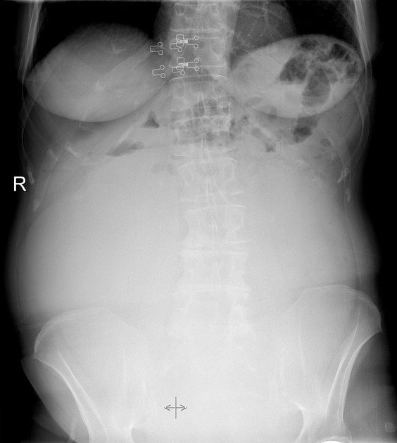
High density mass in central and lower part of abdomen and pelvis. It is sharply demarcated and displaces the bowel loops superiorly.
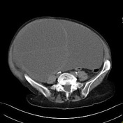


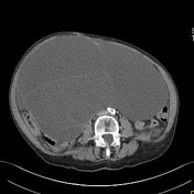
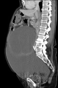
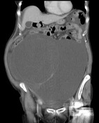
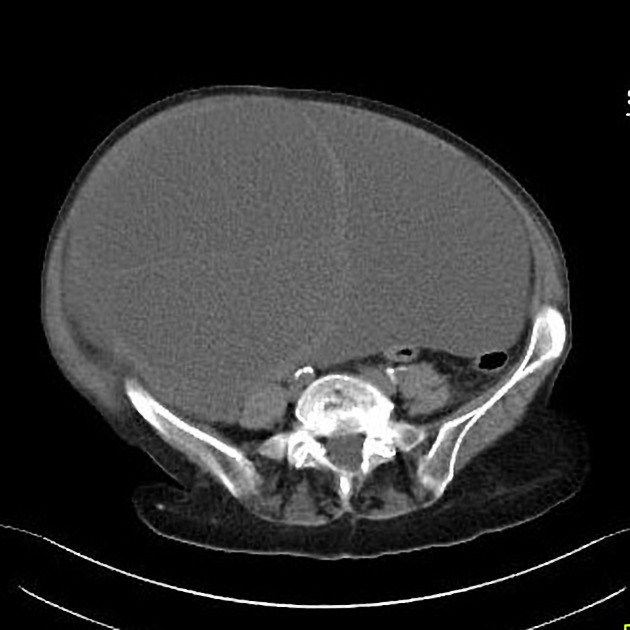
Giant septated cystic mass in pelvis and lower half of abdomen. It does not contain any solid components or calcifications. The walls and septa are mostly thin and sharp. In the lower right part of the cyst there is an enhanced oval structure, probably small uterus with myoma.
Mass effect on surrounding structures is significant, with compressed VCI. Bowel is displaced laterally, with no signs of ileus. There is only mild ascites.
There are no signs of pathologic lymph nodes or metastases.
Contributed: Dr Petra Mrkša
Case Discussion
Hysterectomy was performed. A smooth 6 kg cystic tumor was excised.
Summary of histologic report: giant serous cystadenoma of the right ovary, small uterus with leiomyoma and left ovary that contained simple cyst of size 4 cm.


 Unable to process the form. Check for errors and try again.