
Presentation
Gradual deterioration of left eye vision
Patient Data
Age: 40 years
Gender: Male
From the case:
Primary uveal melanoma

Download
Info
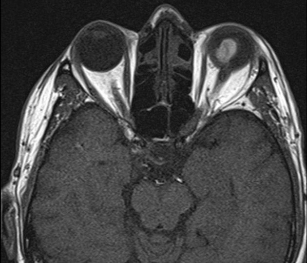
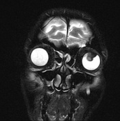
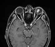
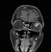
Well defined sharply marginated fungating soft tissue lesion arising from the uveal coating of the superior quadrant of the left eye globe. It displays bright signal on T1, low signal on T2 with avid enhancement on post contrast study. No extra ocular extension and with clear retro orbital fat.
From the case:
Primary uveal melanoma
Download
Info
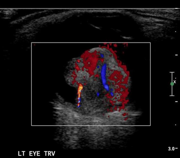
Ocular ultrasound reveals a well defined soft tissue choroidal mass with moderate echogenicity and internal vascularity.
Case Discussion
MRI signal intensities are characteristic of primary uveal melanoma. US with Doppler integration show the characteristic mushroom shape fungating mass with internal vascularity.


 Unable to process the form. Check for errors and try again.