
Presentation
Admitted with falls and cerebellar signs (reduced co-ordination, right lateral nystagmus). Known small vessel disease and previous TIAs, last imaged 5 years ago.
Patient Data
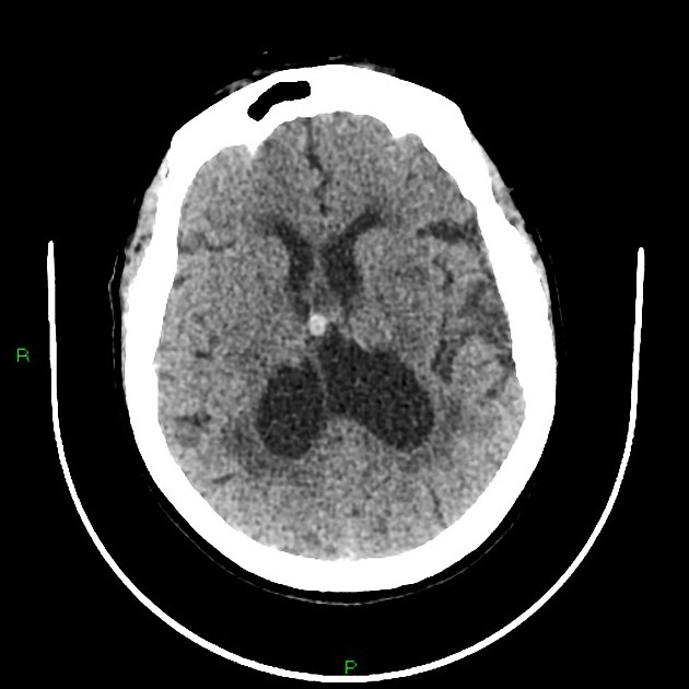
Moderate atrophy.
Marked periventricular and deep white matter ischemic changes.
No acute infarct or hemorrhage. No extra-axial collections or hydrocephalus.
7mm hyperdense cystic structure over Foramen of Monro unchanged from previous imaging - likely colloid cyst.
Small aneurysms of proximal basilar artery and left MCA bifurcation.
Case Discussion
This is an example of chronic small vessel disease, which also includes small unruptured aneurysms and a colloid cyst.
The colloid cyst has remained unchanged on serial CTs over a decade. MPR views in this case were degraded but show it in a position above the foramen of Monro.
Although MRI would be a better modality to determine if there have been any new lesions, particularly in the posterior fossa, the patient was already on stroke preventative medication and this was all attributed to pre-existant small vessel disease.


 Unable to process the form. Check for errors and try again.