
Presentation
Presented to the ER with fever and a previous history of painless growing tumor in neck for two years.
Patient Data
Age: 13 years
Gender: Male
From the case:
Hydatid cyst of the neck
Download
Info
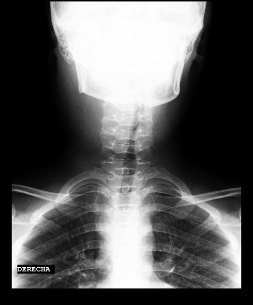
Slight asymmetry of the cervical soft tissues and tracheal deviation to the left.
From the case:
Hydatid cyst of the neck


Download
Info
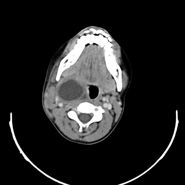
Large cystic mass in the right neck with thick walls and mass effect over trachea and vessels.
Download
Info

An enhanced CT seven months later, shows detachment of membranes, a typical finding of hydatid cyst.
Case Discussion
The neck is an unusual location for hydatid cysts but it has to be taken into account as part of the differential diagnosis for cystic neck lesions in endemic regions.


 Unable to process the form. Check for errors and try again.