
Presentation
Increasingly short of breath on exertion
Patient Data

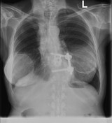
Volume loss in the right upper lobe and increased density and thickening of the paratracheal stripe.
This is an AP projection, however these signs are still visible.
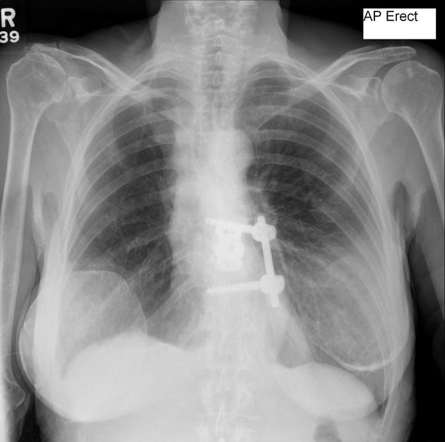
The patient was admitted for bronchial stenting and the post bronchial stenting film is included for interest.
Calcified breast shadows bilaterally.


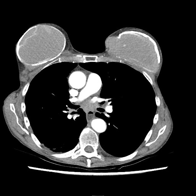
CT study reveals tumor encasing the right main bronchus, with invasion of the right upper lobe bronchus and resultant collapse of the medial segments of the right upper lobe.
The coronal lungs windows illustrate the bronchial collapse and right upper lobe bronchus invasion well.
Heavily calcified host capsules around the prostheses.
Case Discussion
Despite being an AP erect chest x-ray, analysis of the mediastinum is important.
Segmental right upper lobe collapse secondary to lung cancer which invades the right upper lobe bronchus.


 Unable to process the form. Check for errors and try again.