
Radiation pulmonary fibrosis and superior vena caval obstruction
Diagnosis almost certain
Presentation
Known lung cancer received radiotherapy.
Patient Data
Age: 65 years
Gender: Male

Download
Info

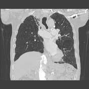
Right upper lobe lung cancer, status post mass resection and radiation therapy, shows right apical architectural distortion, fibrosis, ground glass opacity and consolidation that conforms to the radiation portal.
Bilateral paramediastinal soft tissue fullness associated with SVC obstruction from radiation.
Download
Info
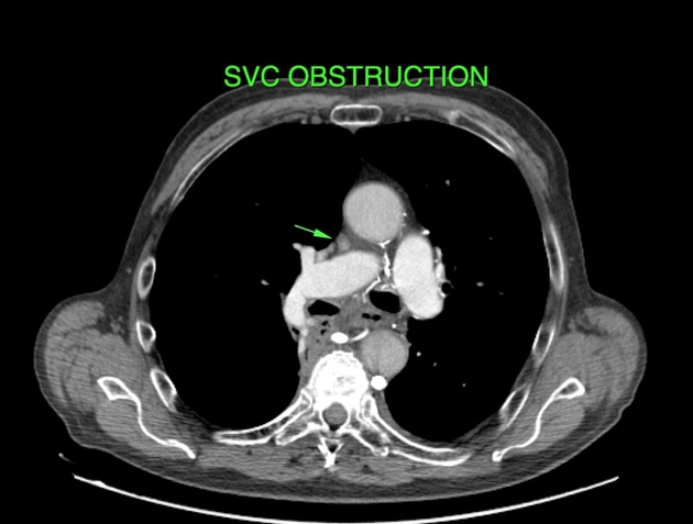
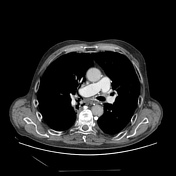
SVC obstruction, narrow lumen with lack of enhancement.
Case Discussion
Radiation pneumonitis implies a ground glass opacity and/or consolidation in context of a clinical pneumonia, whilst in radiation fibrosis is architectural distortion, traction bronchiectasis and fibrosis. This case shows both interstitial pneumonitis and interstitial fibrosis associated with SVC obstruction.


 Unable to process the form. Check for errors and try again.