
Presentation
One year history of painful abdominal wall swelling.
Patient Data
Age: 35 years
Gender: Female
From the case:
Desmoid tumor - rectus abdominis muscle
Download
Info
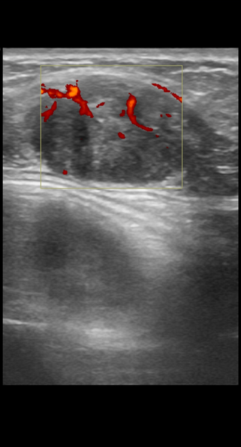
Oval hypoechoic lesion of 3 cm with signal flow in the left rectus abdominis muscle.
From the case:
Desmoid tumor - rectus abdominis muscle


Download
Info
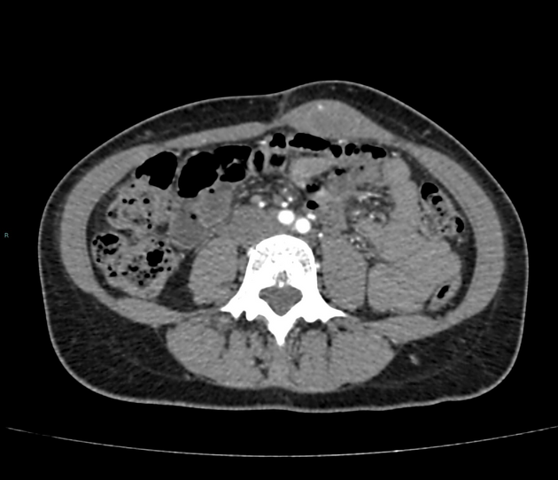
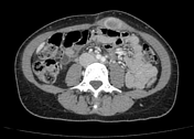
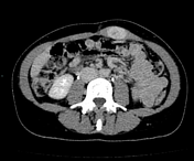
Solid lesion of 3 cm regular margins, with progressive enhancement in the left rectus abdominis muscle mass.
Case Discussion
The lesion was surgically removed.
Histological examination: abdominal fibromatosis: so called desmoid tumor.


 Unable to process the form. Check for errors and try again.