Presentation
Intractable seizures
Patient Data
![](https://prod-images-static.radiopaedia.org/images/15260270/fb569d8ab73a114ee1adf7fa352cc8_thumb.jpeg)
![This study is a stack](/packs/stack-YQKLCKBI.gif)
![](https://prod-images-static.radiopaedia.org/images/15265135/842db90c4e51c9e657bb3b90479874_thumb.jpeg)
![This study is a stack](/packs/stack-YQKLCKBI.gif)
![](https://prod-images-static.radiopaedia.org/images/15268915/688d3d26d4027c72c0c792ed0bfd7b_thumb.jpeg)
![This study is a stack](/packs/stack-YQKLCKBI.gif)
![](https://prod-images-static.radiopaedia.org/images/15266774/bce98d297b5b511586a329986f3fd5_thumb.jpeg)
![This study is a stack](/packs/stack-YQKLCKBI.gif)
![](https://prod-images-static.radiopaedia.org/images/15265590/bf0507648eab1aeb38c8760edf9ea1_thumb.jpeg)
![This study is a stack](/packs/stack-YQKLCKBI.gif)
![](https://prod-images-static.radiopaedia.org/images/15266089/0e156fe777504bcc2a30e7e4ccc387_thumb.jpeg)
![This study is a stack](/packs/stack-YQKLCKBI.gif)
![](https://prod-images-static.radiopaedia.org/images/15264736/23a1c2b2b7b4df777a9d95d6032464_thumb.jpeg)
![This study is a stack](/packs/stack-YQKLCKBI.gif)
![](https://prod-images-static.radiopaedia.org/images/15259028/f1274e421462b474322e61f92061b7_thumb.jpeg)
![This study is a stack](/packs/stack-YQKLCKBI.gif)
![](https://prod-images-static.radiopaedia.org/images/15260943/2fab9e2029042564fea1cd957a25e6_thumb.jpeg)
![This study is a stack](/packs/stack-YQKLCKBI.gif)
![](https://prod-images-static.radiopaedia.org/images/15262485/5516221ac33f0a771ab14f9e677801_thumb.jpeg)
![This study is a stack](/packs/stack-YQKLCKBI.gif)
![](https://prod-images-static.radiopaedia.org/images/15263757/0c5e84c99bae8d94e033aafafad88b_thumb.jpeg)
![This study is a stack](/packs/stack-YQKLCKBI.gif)
![](https://prod-images-static.radiopaedia.org/images/15260270/fb569d8ab73a114ee1adf7fa352cc8_big_gallery.jpeg)
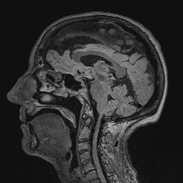
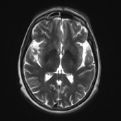
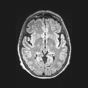
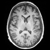
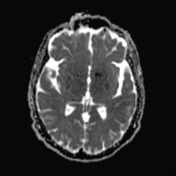
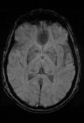
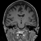
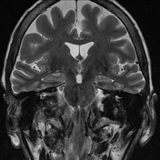
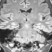
There are two triangular foci increased T2/FLAIR cortical signal in the left posterior frontal lobe involving the precentral gyrus. On the coronal sequences, the apex of the lesion points towards the ventricle with thin linear increased T2/FLAIR extending to the ependymal surface of the left lateral ventricle. This represents the transmantle sign of Blumcke type II focal cortical dysplasia.
There is subsequent loss of normal volume, increased T2/FLAIR signal and loss of normal internal architecture within the left hippocampus in keeping with secondary mesial temporal sclerosis. No forniceal atrophy.
Case Discussion
Great example of focal cortical dysplasia (two lesions) causing mesial temporal sclerosis.