
Presentation
Headache.
Patient Data


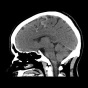

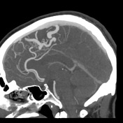
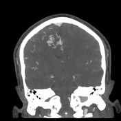
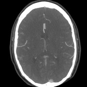
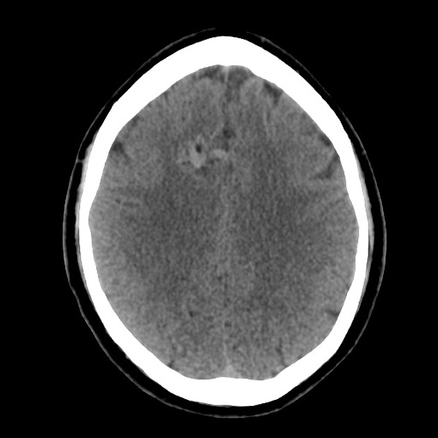
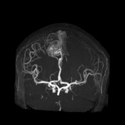
Right frontal paramedian arteriovenous malformation nidus fed predominantly by a branch from the right anterior cerebral artery and drained by enlarged veins that run superiorly through the interhemispheric fissure to the superior sagittal sinus. The nidus measures 2.2 cm in its larger diameter and is not associated with surrounding brain parenchyma intensity abnormalities. Right middle cerebral artery opercular branches are prominent compared to the contralateral circulation and seem to also feed the AVM nidus.
Remainder circle of Willis arteries are unremarkable.
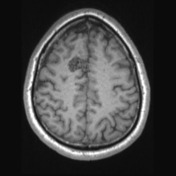
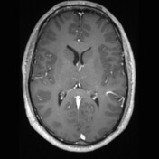
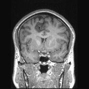
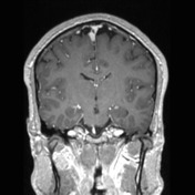
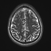
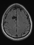
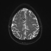
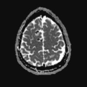
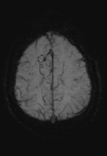
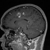
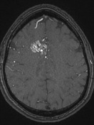


Right frontal paramedian arteriovenous malformation nidus fed predominantly by a branch from the right anterior cerebral artery and drained by enlarged veins that run superiorly through the interhemispheric fissure to the superior sagittal sinus. The nidus measures 2.2 cm in its larger diameter and is not associated with surrounding brain parenchyma signal abnormalities. Right middle cerebral artery opercular branches are prominent compared to the contralateral circulation and seem to also feed the AVM nidus.
Small saccular apparent aneurysm inferiorly oriented is identified on the A1 segment of the right anterior cerebral artery, measuring 3.1 x 2.1 mm.
Remainder circle of Willis arteries are unremarkable.
Remainder appearance and intensity of brain parenchyma are normal.
Ventricular system and cisternal spaces appear normal.
There is no shift of the midline structures.
The visualized orbits and calvarium appear unremarkable. Minima mucosal thickening on the floor of the maxillary sinuses.
Conclusion: Right frontal cerebral arteriovenous malformation (grade 1 accordingly to the Spetzler-Martin AVM grading system). Small probable inferior projection ACA / AComm aneurysm.
Case Discussion
This case illustrates a typical cerebral AVM with a compact nidus and classified as grade 1 accordingly to the Spetzler-Martin AVM grading system.


 Unable to process the form. Check for errors and try again.