
Presentation
Abdominal distension. Hx of bowel surgery 2 years prior.
Patient Data


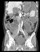
A nasogastric tube is in situ with the tip projected within the anterior gastric body.
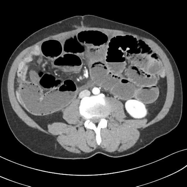
Extensive lobular formations of low attenuation masses (HU 25) surround and encase the liver and spleen scalloping the surfaces. A separate lobular formation extends inferiorly from the level of the IMA to form a larger conglomerate within the pelvis.
There are multiple loops of distended small bowel (largest measuring 40mm diameter) involving the ileum and jejunum. No evidence of intramural gas. The large bowel is predominantly decompressed. No definite transition point can be identified. Bowel staples are noted in the right iliac fossa in keeping with previous resection.
The visualized parenchyma of the liver, spleen, kidneys, adrenal glands and pancreas appear normal. IDC in a collapse bladder. Minor right basal atelectasis is present.
Conclusion
Small bowel obstruction without a definite transition point. This is likely due to adhesions from the extensive lobular formations of fluid density consistent with pseudomyxoma peritonei.
Case Discussion
The patient had a history of bowel resection for appendiceal mucinous adenocarinoma.


 Unable to process the form. Check for errors and try again.