
Presentation
Agitated and confused with a reduced Glasgow Coma Scale (GCS 12). He is pyrexial with a nonspecific history of recent foreign travel. His blood results showed hyponatremia and abnormal liver function tests. In addition, he was found to have a left sided hemiparesis, bulbar palsy and 7th nerve palsy.
Patient Data


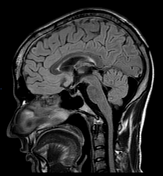
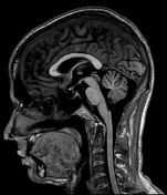
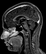
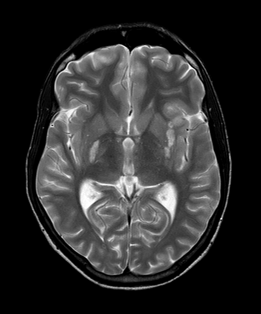
Findings: There are multiple small ring enhancing lesions demonstrated predominantly in the posterior fossa, with at least eight lesions demonstrated in the cerebellum, two lesions in the pons and one lesion left inferomedial temporal lobe. In addition, there are multiple areas of subcortical enhancement throughout both cerebral hemispheres. None of these lesions show restricted diffusion.
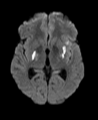
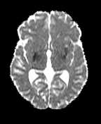
There are large lesions which are not-enhancing, hypointense on T1 and hyperintense on T2/FLAIR, which show restricted diffusion in both corona radiata and the posterior limb of the left internal capsule.
Incidental empty sella.
In view of positive AAFB (acid alcohol fast bacilli) on lumbar puncture: Multiple tuberculomas in the posterior fossa, brainstem and inferomedial left temporal lobe. Evidence of subcortical enhancement elsewhere. Restricted diffusion demonstrated in the deep white matter bilaterally in keeping with ischemia secondary to TB arteritis.
Case Discussion
The combination of radiological and microbiological findings lead to the diagnosis of CNS tuberculosis with multiple tuberculomas and arteritis. The patient was started on a 1-year course of antibiotics (rifampin, isoniazid and ciprofloxacin). Repeat MRI showed the old infarcts and complete resolution of the tuberculomas.


 Unable to process the form. Check for errors and try again.