
Presentation
Chronic hip pain with decreased range of movement. Recent trauma caused the symptoms to aggravate.
Patient Data
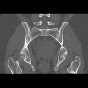

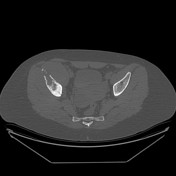

Osteolytic aggressive lesion involving the right iliac bone showing cortical destruction, medullary expansion and periosteal reaction. Associated large soft tissue mass related to the right iliacus muscle.


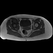
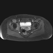
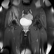
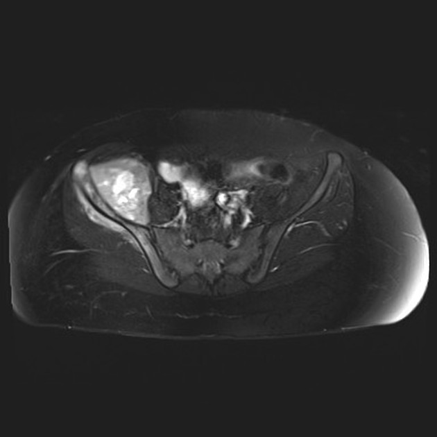
T1 iso to low signal, T2 bright heterogeneous signal together with heterogeneous post contrast enhancement. The related soft tissue component is seen permeating and eroding the iliac bone together with infiltration of the iliacus muscle and to a lesser extent the gluteus minimus muscle.
A simple right ovarian cyst is also seen.
Case Discussion
Typical appearance, age and common site for Ewing sarcoma, showing aggressive behavior with poorly defined margins on CT and radiography, bone destruction, medullary expansion, periosteal reaction and invasion of the surrounding soft tissue. Biopsy proved the diagnosis of Ewing sarcoma.


 Unable to process the form. Check for errors and try again.