
Presentation
Disturbed conscious level, headache and blurring of vision.
Patient Data
Age: 10 years
Gender: Male


Download
Info
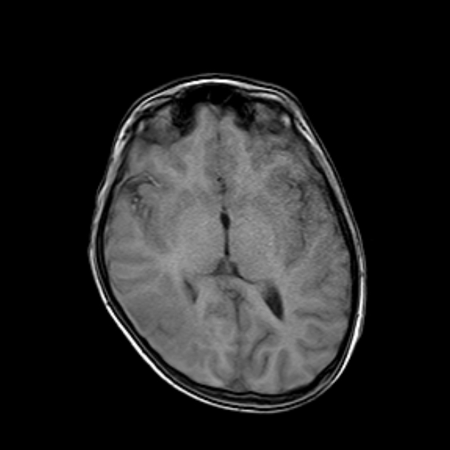
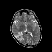
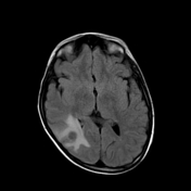
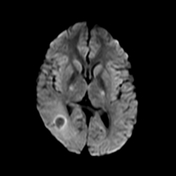
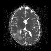
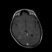
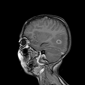
Right occipital space occupying lesion which presents iso signal on T1, hypointense signal on T2/FLAIR with marginal enhancement and surrounded by moderate perifocal edema.
It exerts positive mass effect upon the occipital horn of right lateral ventricle.
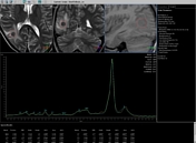
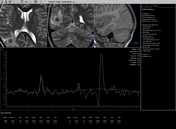
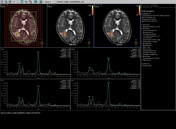
MRS reveals high lipid and lactate levels within the lesion. There is mild NAA reduction and mild Choline rise not consistent with tumorous activity.
Case Discussion
The provisional diagnosis was cerebral tuberculoma which was confirmed by lab investigation and hence the patient improved after anti tuberculous treatment.


 Unable to process the form. Check for errors and try again.